


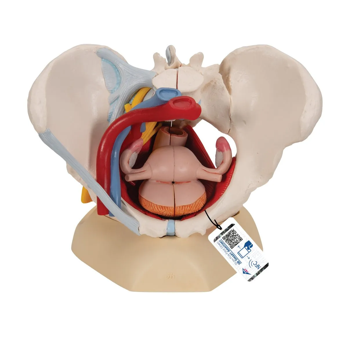
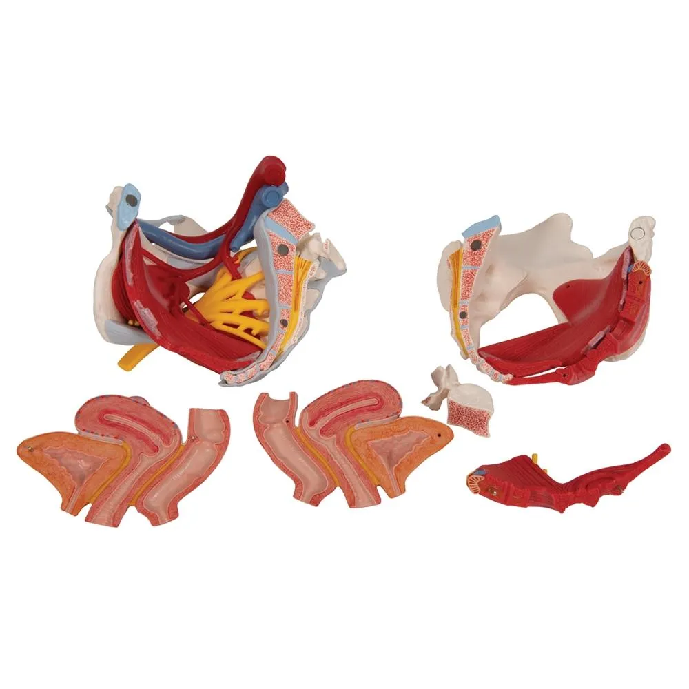

Female Pelvis Model with Ligaments, Vessels, Nerves, Pelvic Floor and Organs, 6 part - Includes 3B Smart Anatomy
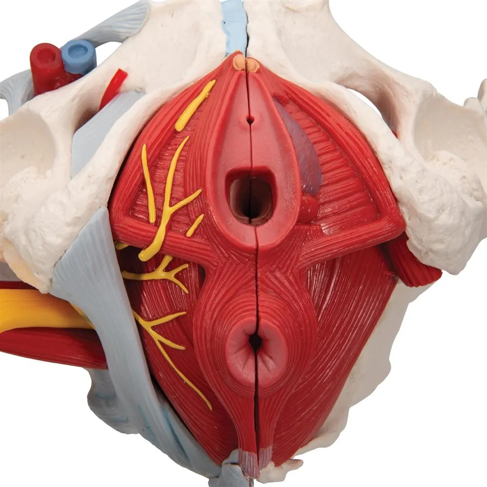
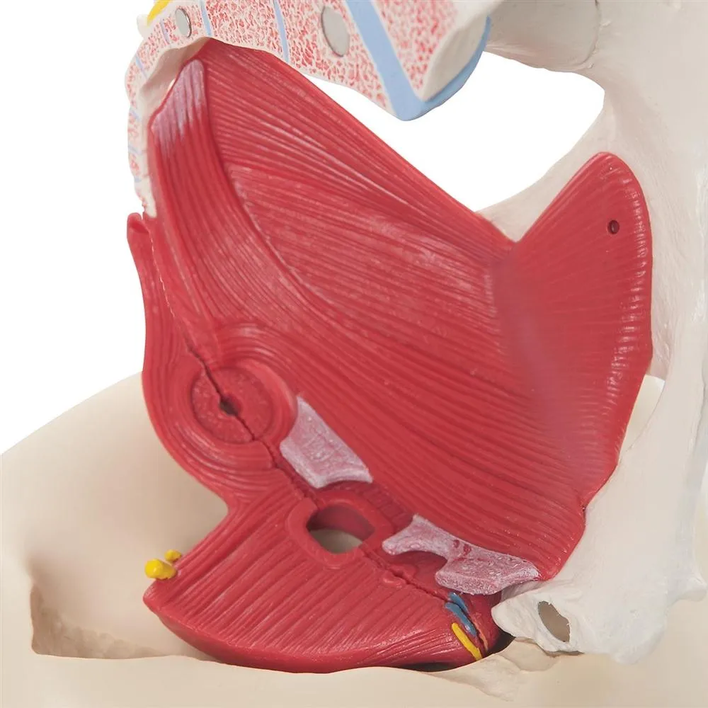
This life size six part model of a female pelvis represents detailed
information about the topography of bones, ligaments, vessels, nerves,
pelvic floor muscles and female genital organs. It presents the whole
pelvic floor with partially removable midsagitally sectioned external
anal sphincter, external urethral sphincter, deep and superficial
transverse perineal and bulbospongiosus.
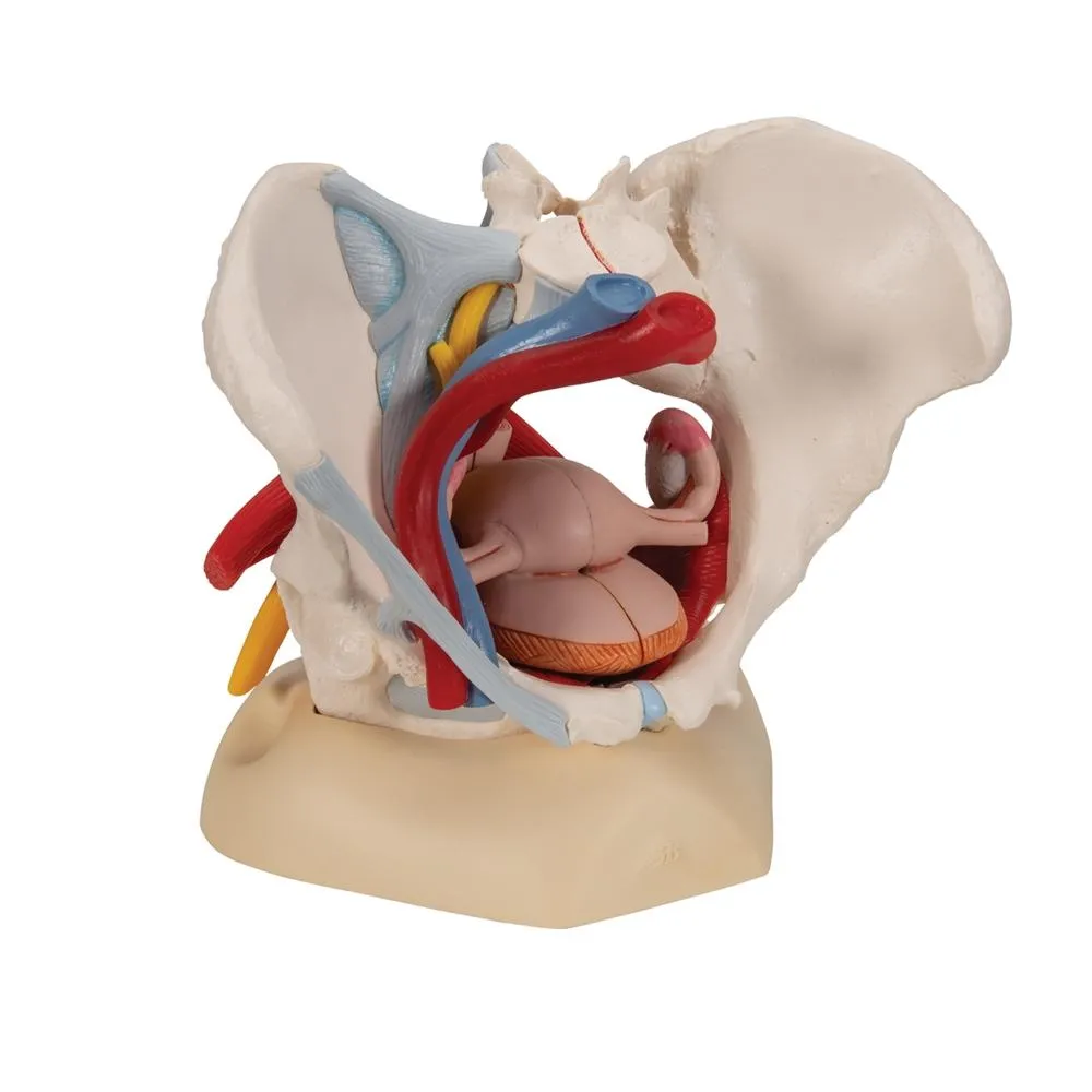
The rectum, uterus with fallopian tubes, ovaries and vagina are also
removable and can be disassembled into both halves by midsagital
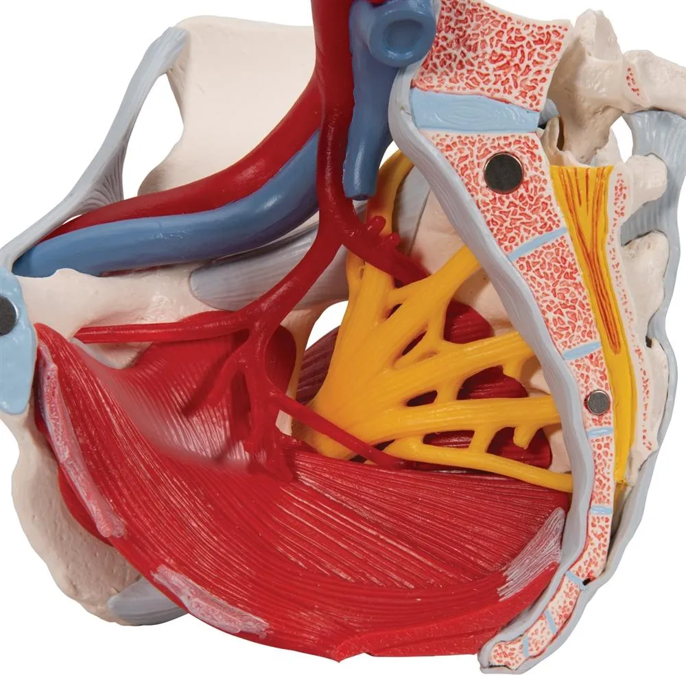
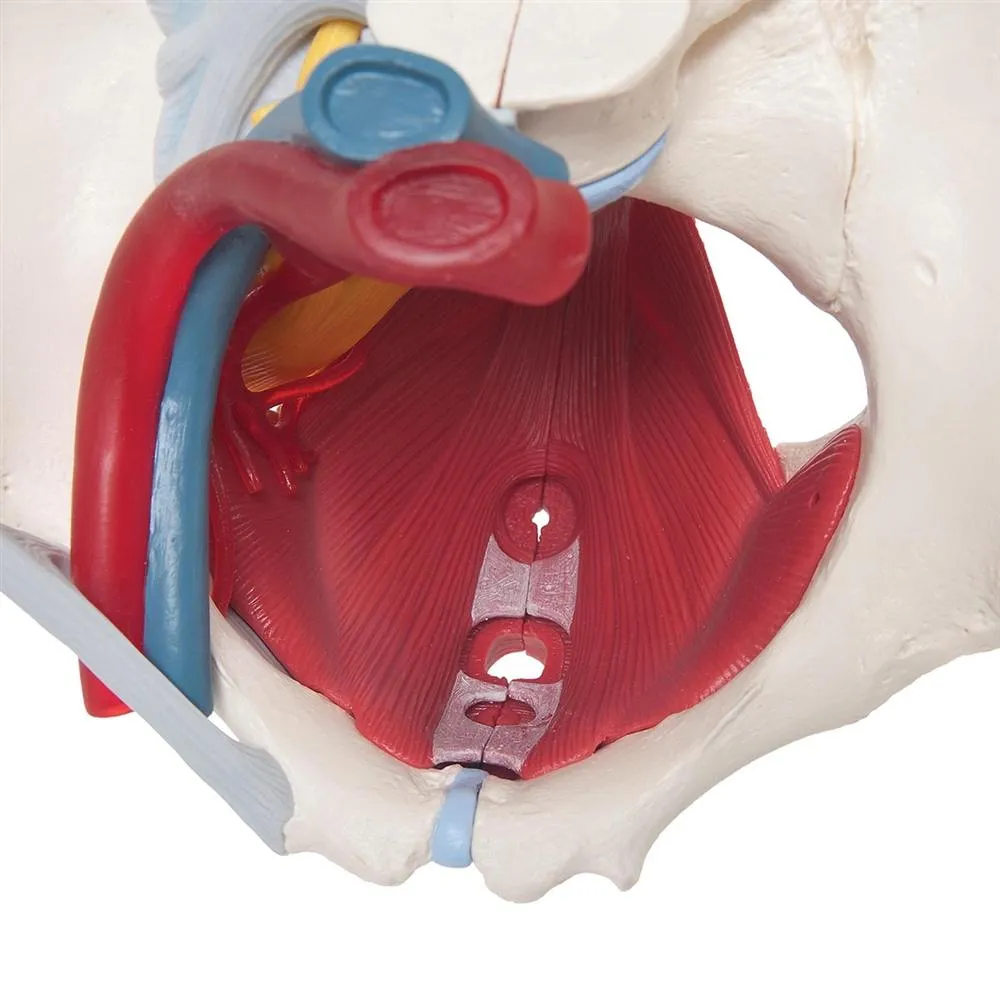
section. The right pelvic half demonstrates the divisions and
topographical anatomy of the common iliac artery, the external and
internal artery and also of the common iliac vein and the external iliac
vein. The right sacral plexus, right sciatic nerve and right pudendal
nerve are also shown.
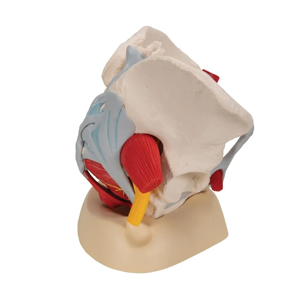
Bones and ligaments presented: Two hip bones, the pubic symphysis, the
sacrum and the coccyx, the fifth lumbar vertebra with intervertebral
disc. A midsagital section through the fifth lumbar vertebra, sacrum and
coccyx, allow both halves of the pelvis to be disassembled revealing a
part of the cauda equina in the vertebral canal. The left half of the
fifth lumbar vertebral body is removable. The right half of the model
shows the following pelvic ligaments: inguinal ligament, sacrotuberous
ligament, sacrospinous ligament, anterior sacroiliac ligaments,
iliolumbar ligament, anterior longitudinal ligament, interosseous
sacroiliac ligament, posterior sacroiliac ligament and obturator.
This model is great for detailed study of the female genital and pelvic anatomy.
GTSimulators by Global Technologies
3B Scientific Authorized Dealer.


")


 - Kristall Strass HT")


